What Does A Animal Cell Look Like Under A Microscope : Animal Cell Under Microscope Structure And Anatomy - When the photos are colorized they look like masterpieces of art.
What Does A Animal Cell Look Like Under A Microscope : Animal Cell Under Microscope Structure And Anatomy - When the photos are colorized they look like masterpieces of art.. What happened to the image in the microscope? The huge variety of cells that have evolved to fulfill different purposes do not always have all the same organelles or structures, but in general terms. It is involved in cell processes,like secretion, plasma membrane repair, cell signaling, and energy metabolism. I cant find any mention of an el08 cell line in the literature. They are trying to locate the cell's dna, which is found in its chromosomes.
Be careful pushing it under the clips that the cover slide doesn't move or crack. When the photos are colorized they look like masterpieces of art. I would like to target insects and mammal tissue. Not every cell is the same. You need to be more precise:
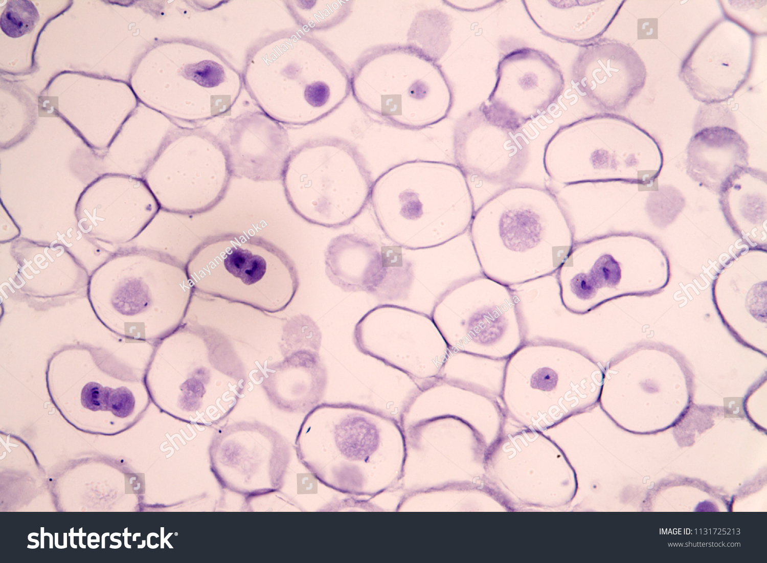
Meiosis Animal Cell Under Microscope Education Stock Photo Edit Now 1131725213 from image.shutterstock.com The procedure can be found in the student guide in the instructions for part 1: Albeit the detail will be minimal without a contrast mechanism or staining or such. In this chapter we will learn about the basic units of life which robert hooke was the first to use the term 'cell' when he studied thin slices of cork with a microscope. I cant find any mention of an el08 cell line in the literature. Animal cells can't do that, although they do have other traits that are commonly shared. Most cells, both animal and plant, range in size between 1 and 100 micrometers and are thus visible only with the aid of a microscope. When looking under a microscope, the cell wall is an easy way to distinguish plant cells. Under a microscope, plant cells from the same source will have a.
In this chapter we will learn about the basic units of life which robert hooke was the first to use the term 'cell' when he studied thin slices of cork with a microscope.
The cell wall looks quite like rectangles jot up. If not, what structures are visible under the staining is used to highlight important features of the tissue as well as to enhance the visualization of the cell or certain cellular components under a microscope. Practice making a wet mount slide of your own cheek cells. Animal cells have unique features that distinguish them from plant and fungi cells. Stem cells in bone marrow divide and transform into blood cells. There are other types of light microscopy in use, but as you start out learning about microscopes cell biologists can see animal and plant cells up close and get a very good idea of what is happening in each cell. They are trying to locate the cell's dna, which is found in its chromosomes. A cell is the smallest unit of life. Cells act like the bricks or building blocks that maintain all of the body's nerves, muscles, tissues, organs, and when looking at an animal cell under a microscope, the thing you first see is the nucleus. Where should scott look to find the chromosomes? To look at a cell close up we need a microscope. It is a sack of water that holds in the chemicals that make up life. Not every cell is the same.
When looking under a microscope, the cell wall is an easy way to distinguish plant cells. Stem cells in bone marrow divide and transform into blood cells. Animal cells can't do that, although they do have other traits that are commonly shared. All living things are made up of smaller structures called cells. Under a light microscope, the cell membrane, nucleus and cytoplasm of a cheek cell (animal cell) you will know if a cell if a plant or animal by looking under a microscope.
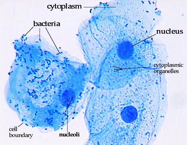
How These 26 Things Look Like Under The Microscope With Diagrams from microbenotes.com Onion cell is a plant cell while human cheek cell is an animal cell. That looks like a secondary electron image to me. If not, what structures are visible under the staining is used to highlight important features of the tissue as well as to enhance the visualization of the cell or certain cellular components under a microscope. It is involved in cell processes,like secretion, plasma membrane repair, cell signaling, and energy metabolism. It has a cell wall, cell membrane, cytoplasm, nucleus cell wall serves as a protective barrier but, it allows the transmission of chemical signals and cellular excretions. You know what, the onion cells look like bricks of a parapet wall when you see it under the low power of this means a cell does various tasks that it was designed to accomplish. You need to be more precise: It doesn't look like that under the microscope, it would have been in black and white.
Learn microscope basics and how the various parts of a microscope work together.
Animal cells include a huge variety of different types of cells. Why do plant cells have more consistent shapes than animal cells? Onion cell is a plant cell while human cheek cell is an animal cell. Under a light microscope, the cell membrane, nucleus and cytoplasm of a cheek cell (animal cell) you will know if a cell if a plant or animal by looking under a microscope. Stem cells in bone marrow divide and transform into blood cells. You know what, the onion cells look like bricks of a parapet wall when you see it under the low power of this means a cell does various tasks that it was designed to accomplish. Select the lowest power objective lens. In fact, under a microscope, a plant cell and an animal cell might seem so similar, in some cases you'd really have to know what you're looking at to tell the difference between if plants and animals are so similar on a cellular level, why do they seem so different when you take a couple steps back? It has a cell wall, cell membrane, cytoplasm, nucleus cell wall serves as a protective barrier but, it allows the transmission of chemical signals and cellular excretions. You need to be more precise: All living things are made up of smaller structures called cells. Cells act like the bricks or building blocks that maintain all of the body's nerves, muscles, tissues, organs, and when looking at an animal cell under a microscope, the thing you first see is the nucleus. You are going to prepare some cells from the inside of your cheek to be the specimen you look at under the microscope.
Stem cells in bone marrow divide and transform into blood cells. Cells consist of cytoplasm enclosed within a membrane, which contains many biomolecules such as proteins and nucleic acids.2 most plant and animal cells are only visible under a light microscope, with dimensions between 1 and 100 micrometres.3 electron microscopy gives a much higher. When the photos are colorized they look like masterpieces of art. Using the microscope to investigate cells. Do the cells in the microscope look like the illustrations shown in this lesson?
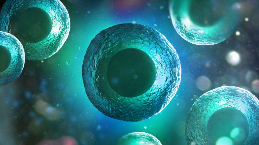
Mini Organs Organoids Rendering Illustration Human Animal Cell Under Microscope 3d 693135160 Singularity Hub from singularityhub.com Animal cells include a huge variety of different types of cells. Using the microscope to investigate cells. What does it mean to be microscopic? I cant find any mention of an el08 cell line in the literature. You know what, the onion cells look like bricks of a parapet wall when you see it under the low power of this means a cell does various tasks that it was designed to accomplish. Practice making a wet mount slide of your own cheek cells. I would be very grateful for info in magnification ranges that i need and maybe some popular models of microscopes. What do plant cells look like under a microscope?
Animal cells have unique features that distinguish them from plant and fungi cells.
Using the microscope to investigate cells. Cells act like the bricks or building blocks that maintain all of the body's nerves, muscles, tissues, organs, and when looking at an animal cell under a microscope, the thing you first see is the nucleus. It doesn't look like that under the microscope, it would have been in black and white. Practice making a wet mount slide of your own cheek cells. With light microscopy i can simply scrape some cells from my cheek smear them on a slide and look at them. The cell wall looks quite like rectangles jot up. With microscopes, we can look directly at some of the objects and processes that are too small to below the microscopic scale. Under a microscope, plant cells from the same source will have a. All living things are made up of smaller structures called cells. Cells consist of cytoplasm enclosed within a membrane, which contains many biomolecules such as proteins and nucleic acids.2 most plant and animal cells are only visible under a light microscope, with dimensions between 1 and 100 micrometres.3 electron microscopy gives a much higher. What is inside a cell and are plant and animal cells the same? Do the cells in the microscope look like the illustrations shown in this lesson? Onion cell is a plant cell while human cheek cell is an animal cell.


0 Comments